Fiber optic array biosensors
Citation
The provided source does not contain a complete citation for the article "Fiber Optic Array Biosensors".
The author is David R. Walt from Tufts University in Medford, MA, USA. The document contains a list of references citing works by Walt or co-authored by Walt, but it does not specify which reference is for the article itself.
Keywords
- Fiber Optic Arrays
- Microwells
- Bead-Based Sensors
- DNA Analysis
- Single-Cell Analysis
- Cell Migration
- Single-Molecule Detection
- Optical Trapping
- Multiplexed Measurements
Brief
There is no information about the content of the article "Analysis of differential gene expression by bead-based fiber-optic array in growth-hormone-secreting pituitary adenomas" in the provided source, so a one-line summary cannot be provided. The provided source describes how optical fiber arrays can be used in biosensing.
Summary
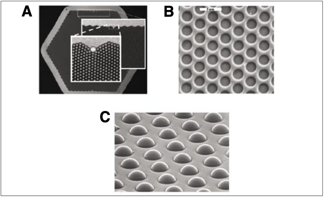
Optical fiber arrays are highlighted for their use in creating high-density biosensors. The arrays are made up of thousands of glass fibers bundled together, each acting as an independent light channel. By etching the fiber cores, microwells are created that can hold beads, cells, or even single molecules. This allows for a wide range of applications:
- DNA Analysis: Beads with attached DNA sequences are loaded into the wells. Different bead types are identified by color or hybridization reactions, enabling high-density genotyping and gene expression analysis.
- Single Cell Analysis: Wells can be sized to hold individual cells, enabling real-time monitoring of cell responses to stimuli or toxins. Different cell types can be included in a single array using fluorescent labels.
- Cell Migration Studies: The fiber surface can be coated with proteins that promote cell adhesion. By tracking fluorescently labeled cells, researchers can rapidly screen the effects of compounds on cell migration.
- Single Molecule Detection: Microwells can be sealed to confine single enzyme molecules. The enzyme's activity can be measured by adding a fluorogenic substrate, which generates a detectable signal only in wells containing the enzyme.
- Optical Trapping: Microbeads in the wells act as lenses, focusing light to create "optical traps." These traps can hold particles like cells or beads, creating temporary "virtual" arrays for various assays.
Key advantages of optical fiber arrays include their availability, high-density format, and versatility in incorporating different materials. The ability to easily modify sensor composition by adding new beads or cells further enhances their utility.