Digital X-ray Sensor
Citation
Keywords
- X-ray imaging
- Digital sensors
- CMOS sensors
- Dental X-rays
- Computed tomography (CT)
- Photostimulable phosphor (PSP) plates
- Scintillation
- Image intensifier screen
Brief
The article provides an overview of X-ray imaging technology, from its historical origins to modern digital sensors, including how CMOS sensors function and their use in dental X-rays.
Summary
Richard Wotiz's January 2012 "Circuit Cellar" article, "Digital X-Ray Sensors," traces the evolution of X-ray technology from its inception to its modern digital applications. The article starts by looking back at Wilhelm Roentgen's 1895 discovery of X-rays and the subsequent use of photographic film and intensifier screens to capture images. Wotiz then describes the emergence of computed tomography (CT) in the 1970s, which uses an X-ray tube and electronic detectors to create 3D images.
The article then shifts its focus to digital X-ray sensors, particularly those used in dentistry. Wotiz highlights the development of digital intraoral X-ray sensors in the 1980s and notes their increasing acceptance as a replacement for traditional film. He explains how these sensors work, describing the role of CMOS sensors, scintillation layers, fiber-optic plates, and the process of capturing and processing the image data.
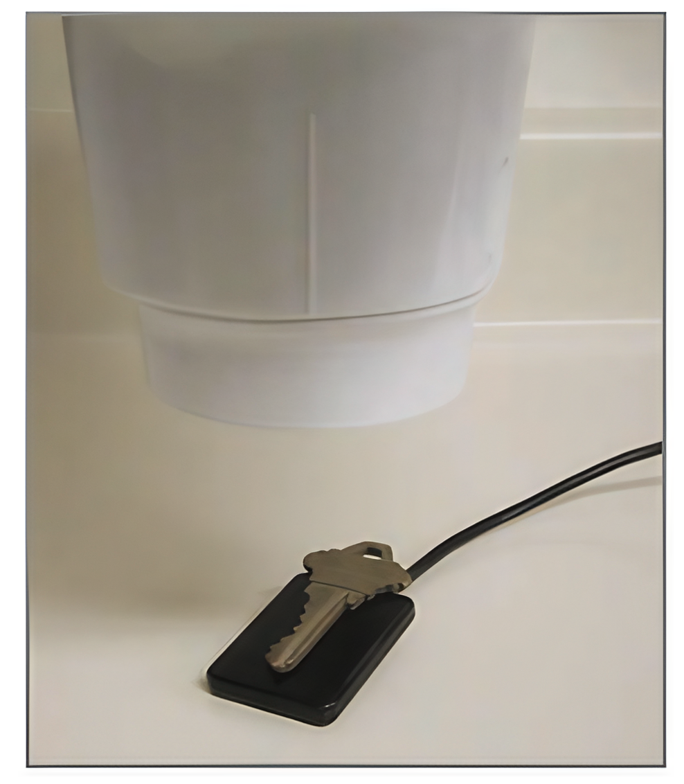
Wotiz also examines a digital X-ray sensor, detailing its components and construction. He provides insights into the sensor's aluminum shield, CMOS imager, scintillation layer, fiber-optic plate, and circuit board. The article concludes with a demonstration of the sensor's capabilities, capturing an image of a brass key. Wotiz discusses the factors that influence image quality, such as tube current, generator voltage, exposure time, and the principle of ALARA.
Origin: https://circuitcellar.com/wp-content/uploads/2012/06/CC2012010601.pdf